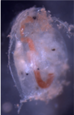
Coral toxicology at Virginia Commonwealth University
In the Lewinski Lab at VCU, we are interested in understanding the interactions of engineered nanoparticles with biological systems. Unlike small molecules, nanoparticles can have a physical interaction as well as a chemical interaction with cells and organisms. This physical interaction was observed between CdSe/ZnS quantum dots and Daphnia magna (Lewinski et al. ES&T 2008), where “sticky” particles adhered to the carapace (exoskeleton) and weighed the organisms down plus adhered to the setae (filter feeding appendages) and inhibited ingestion of food particles.
In addition, nanoparticles can also act as carriers of other chemicals, where adsorption (or sticking) to the surface of a nanoparticle changes the chemical’s biological absorption (transfer into a cell or tissue). This phenomenon is often referred to as the “Trojan Horse effect.” While these properties can be harnessed for beneficial purposes, such as in medicine, they can sometimes lead to unintended consequences in the environment.
When embarking on this coral project, we naturally gravitated towards questions on how engineered nanoparticles interact with corals. We found close to 30 articles with a little over one third reporting on quantum dots in corals. Surprisingly few articles reported on titanium dioxide or zinc oxide, the active ingredients in mineral sunscreens. By comparison, coral exposure to sunscreen ingredients is a current reality; therefore, in this blog post, we take a look at the literature and review available evidence of sunscreen toxicity in corals. Not only will this help us identify experimental paths towards improving our understanding of the mechanisms of toxicity, but also help us make an informed assessment of Jay Sirois’ (Sirois Sci Tot Envi 2019) critique of the science used by regulators to justify banning the sale of certain sunscreens in Hawaii.
In 2018 state lawmakers in Hawaii passed a bill (SB2571 SD2 HD2 CD1) that bans over the counter sale of sunscreens containing oxybenzone or octinoxate as active ingredients starting on January 1, 2021. This bill was proposed based on a growing number of studies, which included a study reporting on the toxicological effects of oxybenzone in the stony coral Stylophora pistillata (Downs et al. in 2016). In a commentary article, Sirois appealed for all available evidence to be reviewed. Citing five articles (Downs 2016, Danovaro 2008, He 2019a, He 2019b, Tsui 2017), the experimental designs of these studies were critiqued for including “single, unreplicated data points.” Let’s look at these articles in more detail.
Table 1. Summary of data from 5 articles cited in Sirios 2019.
| Chemical | Coral | Concentration | Duration | Endpoints | Results | Reference |
|---|---|---|---|---|---|---|
| benzophenone-3 (oxybenzone) | Acropora spp.(3 per test) | 33 µL/L (6% w/w) | 12 hr intervals up to 96 hr | zooxanthellae released, viral infection | Bleaching initiated at 24 hr, 86% bleached after 48 hr, 83% zooxanthellae released | Danovaro 2008 |
| benzophenone-3 (oxybenzone) | Acropora pulchra (3 per test) | 50 µL/L (6% w/w) | 12 hr intervals up to 96 hr | zooxanthellae released, viral infection | Bleaching initiated at 48 hr, 93% bleached after 96 hr, 84% zooxanthellae released | Danovaro 2008 |
| benzophenone-3 (oxybenzone) | Stylophora pistillata (4 per test) | 0, 0.00001, 0.0001, 0.001, 0.01, 0.1, 1 mM (2.28, 22.8, 228 ppb, 2.28, 22.8, 228 ppm) | 8, 24 hr | chlorophyll fluorescence, DNA damage, cell membrane integrity (trypan blue), CaCO3 (Alizarin red stain) | 8 hr - LOEC = 2.28 ppb (light) 22.8 ppb (dark), LC50 = 3.1 ppm (light) 16.8 ppm (dark), EC50 = 107 ppb (light), 436 ppb (dark); 24 hr - LC50 = 103.8 ppb (light) 873.4 ppb (dark); EC50 = 17 ppb (light), 105 ppb (dark) | Downs 2016 |
| benzophenone-3 (oxybenzone) | Platygyra acuta, Porites sp., Pavona decussate, Favites abdita (3 per test) | 13.2-31.7 ng/L | collected samples from Hong Kong | tissue concentrations | 3.6-5.4 ng/g ww (PA), 8.6-31.8 ng/g ww (P), 2.8-18.8 ng/g ww (PD), 10.4-17.6 ng/g ww (FA) | Tsui 2017 |
| benzophenone-3 (oxybenzone) | Seriatopora caliendrum (4 larvae per test) | 1000 µg/L | 7 days | mortality, settlement, bleaching | 5% mortality, 65+/-21.79% settlement, 5 +/-8.7% pale brown, 95+/-8.7% white | He 2019b |
| benzophenone-3 (oxybenzone) | Seriatopora caliendrum, Pocillopora damicornis (3 nubbins per test) | 1000 µg/L | 7 days | bleaching, zooxanthellae density, polyp retraction, tissue concentrations | 50% bleaching, no change in zooxanthellae density, 16.7% polyp retraction (S. caliendrum), no polyp retraction (P. damicornis), 1.42-67.51 ng/g tissue dw | He 2019b |
| ethylhexylmethoxy-cinnamate (octinoxate) | Acropora spp.(3 per test) | 33 µL/L (6% w/w) | 12 hr intervals up to 96 hr | zooxanthellae released, viral infection | Bleaching initiated at 2 hr, 91% bleached after 24 hr, 86% zooxanthellae released | Danovaro 2008 |
| ethylhexylmethoxy-cinnamate (octinoxate) | Acropora pulchra (3 per test) | 50 µL/L (6% w/w) | 12 hr intervals up to 96 hr | zooxanthellae released, viral infection | Bleaching initiated at 48 hr, 91% bleached after 96 hr, 90% zooxanthellae released | Danovaro 2008 |
| ethylhexylmethoxy-cinnamate (octinoxate) | Seriatopora caliendrum, Pocillopora damicornis (3 nubbins per test) | 1000 µg/L | 7 days | mortality, bleaching, zooxanthellae density, polyp retraction | 1000 ug/L LOEC & 83.3% bleaching (S. caliendrum), total polyp retraction @ 10 ug/L (S. caliendrum), 1000 ug/L (P. damicornis), 16.28-198.32 ng/g tissue dw (S. caliendrum), 17.48-168.31 ng/g tissue dw (P. damicornis), | He 2019a |
Interestingly, each research group studied different (1) coral, (2) high concentration, and (3) (sub)acute time points. These differences make comparisons across studies challenging. The acute toxicities observed were at concentrations of 1-60 mg/L oxybenzone or 1-52.5 mg/L octinoxate. These are significantly higher than those measured in the ocean. Measured oxybenzone concentrations in the ocean range in orders of magnitude from nanograms per liter (13.2-31.7 ng/L (ppt) in Hong Kong (Tsui et al. 2017) & 136 ng/L (ppt) in Waikiki Beach, Hawaii (Mitchmore et al. 2019)) to micrograms per liter (0.8 and 19.2 µg/L (ppb) in Hawaiian sites (Downs et al. 2015); 0.01-2.013 µg/L (ppb) in Folly Beach, South Carolina (Bratkovics et al. 2011); 0.116 µg/L (ppb) to 1.4 mg/L (ppm) in Trunk Bay, U.S. Virgin Islands (Downs et al. 2015); 0.127-3.316 µg/L (ppb) Canary Islands (Sanchez Rodriguez et al. 2015)). Fewer measured octinoxate concentrations in the ocean were found (0.03-0.264 µg/L (ppb) in Folly Beach, South Carolina (Bratkovics et al. 2011)).
Only one of these studies (Downs et al. 2016) conducted dose response testing and measured lowest observable adverse effect levels in the concentration ranges (ppb) measured in the ocean. While there are four other articles (Downs 2014, McCoshum 2016, Mitchelmore 2019, Stien 2019) reporting on sunscreen effects in corals, these articles either do not study the two active ingredients being banned in Hawaii (Downs 2014, Stien 2019), do not specify the coral studied (Mitchelmore 2019) or do not study reef building corals (McCoshum 2016). Therefore, our conclusion is that Sirois’ critique is founded and supports the need for more research to fully understand the impact of sunscreen ingredients on corals.
Now focusing on mineral-based sunscreen active ingredients, titanium dioxide and zinc oxide, we found four studies, included in the table below.
Table 2. Summary of TiO2 and ZnO particle studies in corals.
| Chemical | Coral | Concentration | Duration | Endpoints | Results | Reference |
|---|---|---|---|---|---|---|
| TiO2 (anatase, <25 nm, 99.7% purity, Sigma) | Montastraea faveolata (6 per test) | 0.1, 10 mg/L | 17 days | coral gene expression, heat shock protein 70 gene expression, algae count per cm2 coral tissue, titanium tissue concentrations by ICP-MS | d = 19.1+/-3 nm by TEM, 800-1400 nm by DLS, -9.16 mV zeta potential; 14% (0.1 mg/L) and 25% (10 mg/L) decrease in zooxanthellae, gene expression for HSP70 upregulated at 48 h, average TiO2 NP concentration in tissue 6.19 mg/L for 0.1 mg/L dose and 16.58 mg/L for 10 mg/L dose | Jovanovic 2014 |
| Mn-doped TiO2 (Optisol, Oxonica) | Acropora spp. (3 per test) | 6.3 mg/L | 0, 24, 48 hr | algae count in seawater, algae damage, coral bleaching by imaging, microorganism abundance in seawater | release of zooxanthellae highest after 24-28 hr, 6-7% bleaching for TiO2 | Corinaldesi 2018 |
| Al2O3- and dimethicone coated TiO2 (rutile, 20 nm, Eusolex T2000, Merck) | Acropora spp. (3 per test) | 6.3 mg/L | 0, 24, 48 hr | algae count in seawater, algae damage, coral bleaching by imaging, microorganism abundance in seawater | release of zooxanthellae highest after 0-24 hr, 6-7% bleaching for TiO2 | Corinaldesi 2018 |
| ZnO (uncoated, 50-70 nm, Sigma cod. 544906) | Seriatopora caliendrum (7 per test) | 50, 100, 200 μg/L (0.6, 1.2, 2.5 μM) | 24 hr | Zn2+ release from ZnO NPs, SEM of coral, lipid composition changes, qualitative ZnO NP tissue concentrations by SEM | dose dependent change in lipid profile, decrease in 20:4 GPCs | Tang 2017 |
| ZnO (uncoated, 20-200 nm, NPs >50% total particles, Sigma) | Acropora spp. (3 per test) | 6.3 mg/L | 0, 24, 48 hr | algae count in seawater, algae damage, coral bleaching by imaging, microorganism abundance in seawater | release of zooxanthellae highest after 48 hr, 67% bleaching for ZnO, enrichment in prokaryotes and viruses for ZnO | Corinaldesi 2018 |
| ZnO | Stylophora pistillata (3 per test) | 10-5000 μg/L | 35 days (measurements at 0, 7, 14, 21, 28, 35 days) | photosynthetic efficiency | nubbin death at >=1000 ug/L ZnO, LC50 = 24 ug/L for ZnO at 90 d, Bleaching 50% = 864 ug/L for ZnO at 35 d, ZnO induced greatest damage in PSII photosynthetic activity at lowest concentration 10 ug/L, 38% inhibition at 100 ug/L ZnO | Fel 2019 |
A similar trend was observed in these studies with different (1) coral and (2) high concentrations tested. Interestingly, there were some longer term experiments (17, 35 days) conducted. To put the exposure concentrations in perspective, surface water and water column concentrations measured at 3 beaches in Marseille, France included 100-900 µg/L and 20-50 µg/L TiO2 as well as 10-15 µg/L and 1-3 µg/L ZnO, respectively (Labille et al. 2020). These four studies preliminarily suggest that ZnO is comparatively more toxic than TiO2, although this is based on a single concentration, single group reported experiment. Similarly more research is needed to verify that mineral-based sunscreens are indeed “reef safe” as currently marketed.
Coral reefs are experiencing changes in environmental and anthropogenic factors resulting in increased coral bleaching events. To better understand the mechanisms behind these effects, toxicological research must be performed. Coral toxicology studies are currently hindered due to a lack of robust coral models. While Pocillopora damicornis and Stylophora pistillata are often called “guinea pig” corals since they are well studied, there is a need to develop robust coral cellular and polyp models for studying biological responses and impacts. Our team is actively working on developing coral cellular and polyp models for toxicity testing, and we look forward to sharing our experimental results in our next blog entry.
References
Corinaldesi, C., Marcellini, F., Nepote, E., Damiani, E. and Danovaro, R., 2018. Impact of inorganic UV filters contained in sunscreen products on tropical stony corals (Acropora spp.). Science of The Total Environment, 637, pp.1279-1285.
Danovaro, R., Bongiorni, L., Corinaldesi, C., Giovannelli, D., Damiani, E., Astolfi, P., Greci, L. and Pusceddu, A., 2008. Sunscreens cause coral bleaching by promoting viral infections. Environmental health perspectives, 116(4), pp.441-447.
Downs, C.A., Kramarsky-Winter, E., Fauth, J.E., Segal, R., Bronstein, O., Jeger, R., Lichtenfeld, Y., Woodley, C.M., Pennington, P., Kushmaro, A. and Loya, Y., 2014. Toxicological effects of the sunscreen UV filter, benzophenone-2, on planulae and in vitro cells of the coral, Stylophora pistillata. Ecotoxicology, 23(2), pp.175-191.
Downs, C.A., Kramarsky-Winter, E., Segal, R., Fauth, J., Knutson, S., Bronstein, O., Ciner, F.R., Jeger, R., Lichtenfeld, Y., Woodley, C.M. and Pennington, P., 2016. Toxicopathological effects of the sunscreen UV filter, oxybenzone (benzophenone-3), on coral planulae and cultured primary cells and its environmental contamination in Hawaii and the US Virgin Islands. Archives of environmental contamination and toxicology, 70(2), pp.265-288.
Fel, J.P., Lacherez, C., Bensetra, A., Mezzache, S., Béraud, E., Léonard, M., Allemand, D. and Ferrier-Pagès, C., 2019. Photochemical response of the scleractinian coral Stylophora pistillata to some sunscreen ingredients. Coral Reefs, 38(1), pp.109-122.
He, T., Tsui, M.M.P., Tan, C.J., Ng, K.Y., Guo, F.W., Wang, L.H., Fan, T.Y., Lam, P.K.S. and Murphy, M.B., 2019a. Comparative toxicities of four benzophenone ultraviolet filters to two life stages of two coral species. Science of the Total Environment, 651, pp.2391-2399.
He, T., Tsui, M.M.P., Tan, C.J., Ma, C.Y., Yiu, S.K.F., Wang, L.H., Fan, T.Y., Lam, P.K.S. and Murphy, M.B., 2019b. Toxicological effects of two organic ultraviolet filters and a related commercial sunscreen product in adult corals. Environmental pollution, 245, pp.462-471.
Jovanović, B. and Guzmán, H.M., 2014. Effects of titanium dioxide (TiO2) nanoparticles on caribbean reef‐building coral (Montastraea faveolata). Environmental toxicology and chemistry, 33(6), pp.1346-1353.
Lewinski, N.A., Zhu, H., Jo, H.J., Pham, D., Kamath, R.R., Ouyang, C.R., Vulpe, C.D., Colvin, V.L. and Drezek, R.A., 2010. Quantification of water solubilized CdSe/ZnS quantum dots in Daphnia magna. Environmental Science & Technology, 44(5), pp.1841-1846.
McCoshum, S.M., Schlarb, A.M. and Baum, K.A., 2016. Direct and indirect effects of sunscreen exposure for reef biota. Hydrobiologia, 776(1), pp.139-146.
Mitchelmore, C.L., He, K., Gonsior, M., Hain, E., Heyes, A., Clark, C., Younger, R., Schmitt-Kopplin, P., Feerick, A., Conway, A. and Blaney, L., 2019. Occurrence and distribution of UV-filters and other anthropogenic contaminants in coastal surface water, sediment, and coral tissue from Hawaii. Science of the total environment, 670, pp.398-410.
Sirois, J., 2019. Examine all available evidence before making decisions on sunscreen ingredient bans. Science of The Total Environment, 674, pp.211-212.
Stien, D., Clergeaud, F., Rodrigues, A.M., Lebaron, K., Pillot, R., Romans, P., Fagervold, S. and Lebaron, P., 2018. Metabolomics reveal that octocrylene accumulates in Pocillopora damicornis tissues as fatty acid conjugates and triggers coral cell mitochondrial dysfunction. Analytical chemistry, 91(1), pp.990-995.
Tang, C.H., Lin, C.Y., Lee, S.H. and Wang, W.H., 2017. Membrane lipid profiles of coral responded to zinc oxide nanoparticle-induced perturbations on the cellular membrane. Aquatic Toxicology, 187, pp.72-81.
Tsui, M.M., Lam, J.C., Ng, T.Y., Ang, P.O., Murphy, M.B. and Lam, P.K., 2017. Occurrence, distribution, and fate of organic UV filters in coral communities. Environmental Science & Technology, 51(8), pp.4182-4190.